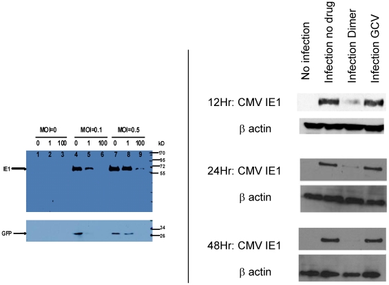
Figure 5. Western blot for IE1, GFP and β actin in artemisinin treated CMV infected cells.
5a: HFF were treated with artemisinin 0, 1 µM and 100 µM and infected with GFP-tagged Towne. Western blot for IE1 and GFP performed at day 5 post infection. 5b: HFF were infected with a clinical isolate, SB, and treated with either dimer primary alcohol (1 µM) or GCV (10 µM). Western blot for IE1 was performed at 12 hr (MOI = 3), 24 hr and 48 hr (MOI = 1) post infection.

