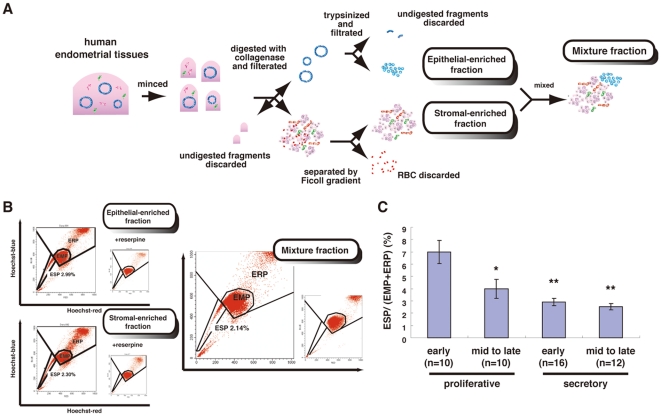
Figure 1. Isolation of ESP and EMP cells.
(A) Summary of procedures for the preparation of epithelial-enriched and stromal-enriched fractions and the mixture of both fractions from human cycling endometria. (B) Flow cytometric distribution of ESP cells, EMP cells, and the endometrial replicative population (ERP) in each of the three fractions stained with Hoechst 33342 indicated in Figure S1. Addition of 50 µM reserpine resulted in the disappearance of the ESP fraction (inset in each panel). (C) Proportion of ESP in the whole fraction dissociated from human endometria at different phases of the menstrual cycle. * P<0.01 and ** P<0.00005, versus early proliferative phase. Each bar indicates the mean ± SEM. n = 48.

