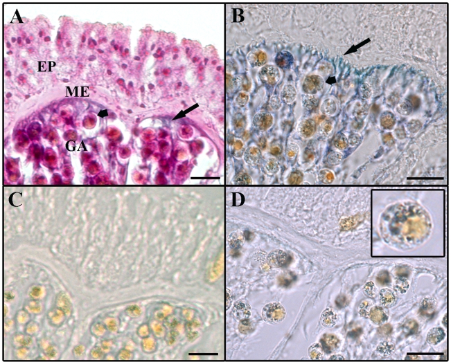
Figure 2. Cross-sections of Lobophytum pauciflorum tissue showing areas of NADPH-diaphorase staining.
A: Section counterstained with H&E. B: NADPH-diaphorase staining evident in host tissue (long arrows) and in symbiotic dinoflagellates (short arrows). C: Negative control without staining; the NOS cofactor NADPH was omitted from the reaction. D: Negative control; the NOS inhibitors L-NMA and L-NAME were added to the staining reaction. NADPH-diaphorase staining is evident in dinoflagellates (insert) but not in host tissue. EP: epidermis, GA: gastrodermis, ME: mesoglea. Scale bars: 20 µm.

