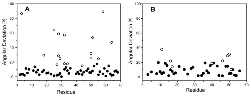
Figure 7.
Residue by residue comparison of the final RSDC vector orientations with the ubiquitin (A) and protein GB1 (B) X-ray structures (1UBQ and 1PGB, respectively). Shown are both the best fit orientations (●) in best agreement with the X-ray structures as well as all other orientations (○) which agree with the experimental RDC data within 3σD.

