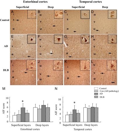
Figure 2.
AIF immunoreactivity (AIF-ir) in the entorhinal and temporal cortices of AD, DLB, control with AD pathology, and control brains. A–L: Immunostaining shows that AIF (brown) was mainly expressed in neurons. Arrows indicate the cytoplasm labeling of AIF in control (A–D), DLB (I–L), as well as the nuclear labeling of AIF in AD (E–H) brains. Insets show higher magnification of neurons indicated by the arrows. Scale bar: 100 μm (A–L). Semiquantitative evaluation of AIF expression in entorhinal (M) and temporal cortices (N) in AD (n = 10), DLB (n = 8), control with AD pathology (n = 8), as well as control brains (n = 11). *P < 0.05 as compared with age-matched control group using Kruskal–Wallis test.

