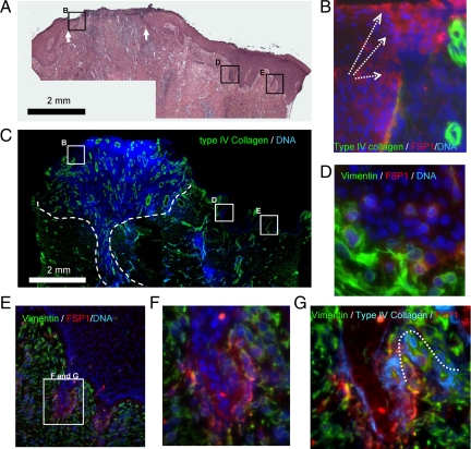
Figure 1.
EMT characteristics in reepithelization. A: Typical reepithelization of cutaneous wound healing of thermal burn patients is illustrated by H&E staining. White arrows indicate the wound bed. The interior box in B, D, and E are designated for the regions of migration tongue and adjacent deep ridges, respectively, and illustrated in B, D, and E, accordingly. B: Epithelial cells in migrating tongues are indicted by their positions above the BMZ (type-IV collagen immunoflorescent staining, green). The trend of migration is highlighted by white dashed arrows. Gain of mesenchymal features by the epithelial cells in the migration tongues is demonstrated by FSP1 expression (IF staining, red). Nuclei DNA are stained by DAPI in blue. C: Granulation (dashed lines), as marked by cell proliferation (DNA, blue) and angiogenesis (IF staining, type-IV collagen, green), is evident in the reepithelization phase. D–F: Gain of mesenchymal features by the epithelial cells in the deep ridges of epidermis adjacent to wound center is demonstrated by FSP1 and vimentin expression. The EMT features are absent in the epithelial cells of spinous and granular layers. G: The wrecked BMZ at the endings of deep ridges is indicated by discontinued IV collagen stained (IF, blue and the dotted line).

