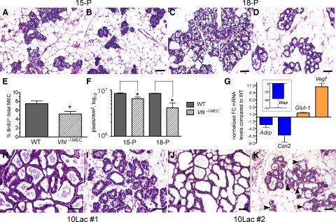
Figure 2.
Reduced alveolar proliferation and differentiation in response to Vhl deletion via MMTV-Cre. Mammary glands were harvested from floxed control (A, C, H, J) or Vhl −/−MEC (B, D, I, K) glands at day 15 (A–B; 15-P, ×200 magnification) or day 18 of pregnancy (C–D; 18-P, ×200 magnification), day 10 of the first lactation (H–I; 10Lac#1) or day 10 of the second lactation (J–K; 10Lac#2), sectioned and stained with H&E. Scale bars = 10 μm. A–B: Note the decreased number of alveoli per field and the lack of differentiation in Vhl −/−MEC glands at 15-P. C–D: Fewer alveoli were also observed in Vhl −/−MEC glands at 18-P. E: The percentage of BrdU+ cells per total number of MEC was determined at day 15-P in individual mice (n = 5/genotype), and the grand mean compared between genotypes. *P < 0.043 F: The number of hematoxylin-positive pixels associated with MEC/mm2 gland scan area was compared using whole-slide scanned, digitized slides by the automated Aperio Pixel Density algorithm as described in the methods. The mean number of pixels plus the SEM is presented. *P < 0.02 G: Real-time PCR was performed to compare expression of Adrp, ß-casein (Csn2), Wap, Glut-1, and Vegf mRNAs in control and Vhl −/−MEC glands harvested at day 18 of pregnancy. The normalized fold-change (FC) in gene expression is expressed relative to expression levels observed in cDNA samples from control glands. Blue or orange bars indicate those genes for which relative expression decreased or increased, respectively. The average fold-change plus the SEM is presented. H–K: H&E-stained sections were prepared from mammary glands harvested from mid-lactation dams during the first (10Lac#1; H–I, ×400 magnification) or second (10Lac#2; J–K, ×200 magnification) cycles of lactation from the same cohort of test and control mice. As expected, the morphology of the wild-type gland remains relatively unchanged from the first to the second lactation (H versus J), whereas large lipid droplets are still present when Vhl is deleted (K; black arrowheads). Digitized images of representative H&E-stained slides from these experiments are available for viewing in the Supplemental Database at http://ajp.amjpathol.org.

