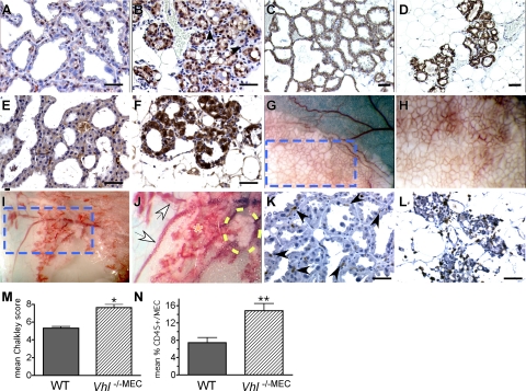
Figure 4.
Changes in HIF-1α, GLUT-1, and VEGF expression, microvessel density and immune cell infiltration in multiparous mice. A–F: Deletion of Vhl in the mammary epithelium results in uniform over-expression of HIF-1α (B), GLUT-1 (D), and VEGF (F) by MEC in thrice multiparous mice. Paraffin sections from formalin-fixed control (WT) or Vhl −/−MEC mammary glands generated from Wap-Cre transgenic stocks were prepared from glands isolated from lactating dams during the third round of lactation. Scale bars = 10 μm. Following antigen retrieval in citrate buffer, sections were stained with antibodies to HIF-1α, GLUT-1, VEGF, or the pan-leukocyte marker CD45. A–B: HIF-1α signal (brown nuclear stain) was detected in approximately 60% of MEC in WT lactating tissue (A), whereas intense nuclear staining as well as cytoplasmic staining of HIF-1α was observed in all MEC in which Vhl was deleted (B). Note the lack of HIF-1α staining in the stromal cells immediately adjacent to and surrounding the alveoli (B); the majority of these stromal cells have the morphology of myoepithelial cells (white arrowheads) or leukocytes (black arrowheads), ×400 magnification. C: As expected, GLUT-1 is expressed by all WT MEC at lactation, but there was an extreme up-regulation of GLUT-1 expression in the Vhl −/−MEC glands (D), ×200 magnification. E–F: Similarly, low levels of VEGF were expressed by WT glands (E), whereas expression of VEGF was strongly up-regulated in the Vhl −/−MEC glands (F), ×400 magnification. G–H: Deletion of Vhl results in hypervascularity and hemorrhage of the mammary gland. The gross appearance of thrice parous wild-type (G–H) or Vhl −/−MEC (I–J) thoracic mammary glands was compared by imaging live, anesthetized mice under a stereozoom dissecting microscope. The edge of the thoracic gland is shown in each low power field (G, I, ×12.5 magnification). The dashed blue box indicates the area highlighted in panels (G) and (I). In these panels individual alveoli and their supporting blood vessels can be visualized (H, J, ×25 magnification). In control dams, all alveoli are fully distended with milk (G, white opaque material), and the uniform basketlike network of vasculature that surrounds each alveolus can be identified (H). In contrast, in Vhl −/−MEC glands, the majority of vessels supporting the alveoli are hyperdilated, overall vessel density appears to be increased and there are areas of hemorrhage (J, orange asterisk). The large intramammary vessels are also dilated (J, white arrows). Moreover, the paucity of milk-containing alveoli in the Vhl null gland is striking, as there are only a few lobules capable of producing milk (yellow dashed oval). K–L: Sections were stained with anti-CD45 antibodies. Very few CD45+ cells were detected in the stroma of wild-type lactating glands (K, arrowheads), whereas there was an increase in the number of CD45+ cells present in the Vhl −/−MEC glands (L). To quantitate changes in microvessel density and leukocyte infiltration, Chalkley analysis was performed following immunostaining for CD34 (M) and the mean percentage of CD45+ cells/total number of MEC was determined (N). The mean Chalkley score and the percentage of CD45+/MEC plus SEM are presented; asterisks indicate statistically significant changes, as measured by an unpaired t-test (M–N). *P = 0.007, **P = 0.02.

