Figure 4.
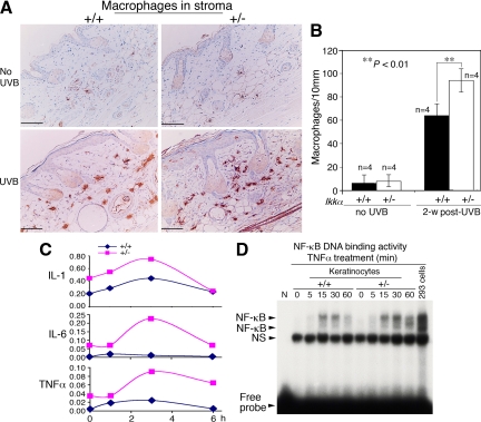
Reduction of IKKα expression elevates inflammation in the skin. A: Detection of macrophages in Ikkα+/+ (+/+) and Ikkα+/− (+/−) paraffin-embedded skin sections, immunohistochemically stained with a macrophage marker. Brown color, macrophages; blue color, nuclear countingstaining with hemetoxylin; UVB, UVB irradiation for 2 weeks. B: Comparison of numbers of macrophages in the skin stroma of UVB-irradiated Ikkα+/+ (+/+) and Ikkα+/− (+/−) mice shown in A. Groups of mice (n = 4) used. 2-w post-UVB, skin prepared from mice at week 2 after UVB irradiation with 100 mJ/cm2. **P < 0.01, t test. Scale bars = 30 μm. C: Relative levels of TNFα, IL-6, and IL-1 mRNA in UVB-irradiated Ikkα+/+ (+/+) and Ikkα+/− (+/−) primary cultured keratinocytes detected using RT-PCR. Levels of GAPDH mRNA were used to normalize the expression levels of these cytokines. Each point (n = 3) used. 2, 4, 6 hours, primary cultured keratinocytes were collected at 2, 4, and 6 hours after irradiation (100 mJ/cm2). D: NF-κB DNA-binding activity in primary cultured keratinocytes treated with TNFα (10 ng/ml) from 0 to 60 minutes. 293 cells treated with TNFα for 30 minutes were used as a positive control. NS indicates nonspecific band.

