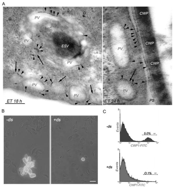
Figure 8. gμ2 plays an essential role during encystations.
(A) Enlarged immunoelectromicrograph of encysting WB1267 trophozoites showing PVs surrounding a dense ESV at 18 h post-encystation. gμ2 is primarily observed in the PVs in patches (red arrowheads). At 24 h of encystation, the CWPs are released from the ESVs and assembled, forming the cyst wall. In addition to the PVs, gμ2 is localized on the inner side of the plasma membrane, coincident with the site of electrodense material accumulation outside the cell. Also note electrodense (CWPs) material in some PVs (arrows). Bars, 0.1 μm. G: electron-dense glycogen deposits. PS: periplasmic space. ET: encysting trophozoite. (B) IFA shows mature cysts in −ds cells but not in +ds cells by the detection of CWP1 in the cyst wall (green). Bar, 10 μm. (C) Flow cytometric analysis demonstrating that the positive population represented by the mature cysts (M1) is decreased in +ds (0.1%, bottom panel) compared with −ds cells (5.0%, top panel). One representative experiment of four performed is shown.

