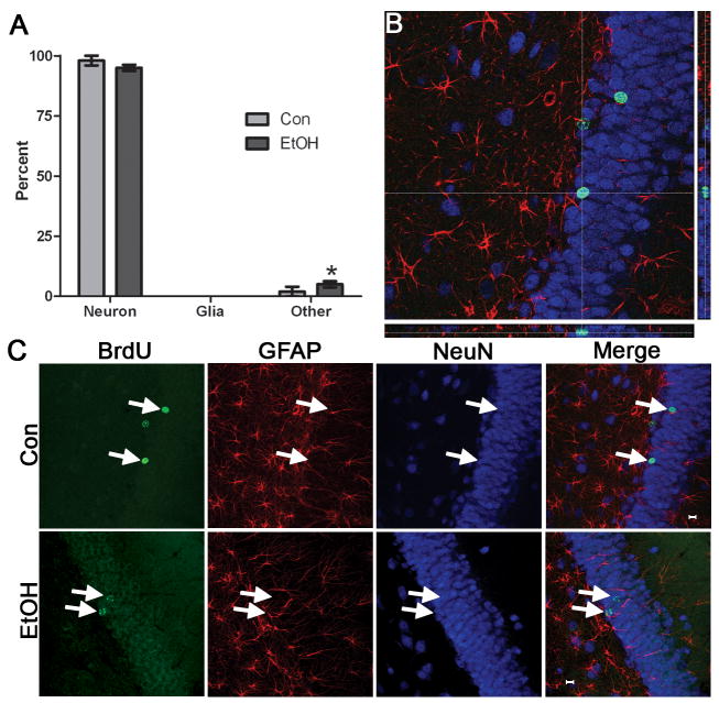
Figure 5.
Binge alcohol administration does not alter the phenotype of cells born during alcohol intoxication. BrdU was injected at 4D and rats were sacrificed 28 days later (4D+28). A) Triple fluorescent labeling for BrdU, a neuron-specific marker (NeuN) and an astroglia specific marker (GFAP) was assessed for colabeling in 50 BrdU+ cells per subject. The percentage of BrdU+ cells that expressed a neuronal phenotype (BrdU+/NeuN+) or an astroglial phenotype (BrdU+/GFAP+) was similar in control (n=5) and alcohol-exposed tissue (n=4). There was only a significant increase in “other” cells (BrdU+/NeuN-/GFAP-) in the alcohol-exposed tissue, which merely shows that other types of cells are proliferating and remaining after binge treatment. These few other cells could be microglia, endothelial cells, or oligodendroglia. A representative orthogonal view of reconstructed Z-stacks is shown in B where BrdU is labeled in green, NeuN in blue, and GFAP in red. The crosshairs are placed atop the cell of interest such that colabeling can be assessed from all planes. C. Representative images of individual fluorochromes showing BrdU+ cells (green), GFAP+ cells (red), NeuN+ cells (blue), and merged in both the control and alcohol-exposed tissue.

