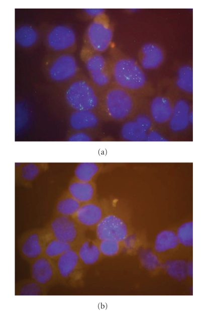
Figure 4.
Interphase FISH assay for the demonstration of EBV in cell lines. Hybridization was performed with EBV cosmid cM-SalI-A labeled with Spectrum Green by nick-translation. Nuclei were stained with DAPI. (a) Nuclei of cell line BONNA-12 contains heterogeneous numbers of EBV copies. (b) Cell line DOHH-2 displays only few cells contaminated with EBV.

