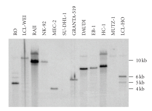
Figure 6.
Southern blot analysis of different cell lines. Fifteen μg genomic DNA of the cell lines were digested with XhoI and the restriction fragments were separated on an agarose gel. The DNA was blotted onto nylon filters and the blot was hybridized to a ca. 900 bp 32P-labeled PCR product spanning the 5′-region of EBV (LMP fragment 1). A rehybridization of the same blot with a probe for the 3′-region of the linear EBV genome after stripping revealed the same bands, indicating that circular EBV DNA is detected.

