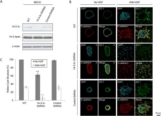
Figure 3.
Loss of 14–3–3σ in MDCK cells results in loss of epithelial polarity. (A) Immunoblot analysis to detect 14–3–3σ and all 14–3–3 protein levels in wild-type (WT), 14–3–3σ–shRNA-expressing, and control shRNA-expressing MDCK cells. β-Actin was used as loading control. (B) Representative immunofluorescent images of aforementioned MDCK cells in 3D collagen cultures, with the exception of pictures having asterisks (*) showing the major abnormal structures. ZO-1 (green) was stained for tight junction, and E-cadherin (red) was stained for adhesion junction. Nuclei were counterstained with DAPI (blue). (C) Percentages of hollow cyst structures in the collagen 3D cultures of the aforementioned MDCK cells without (gray) and with (white) HGF treatment. All experiments were performed in triplicate. Results significantly different from wild-type cells are indicated by asterisks: (*) P < 0.05; (**) P < 0.01.

