Figure 6.
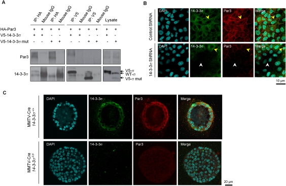
Association of 14–3–3σ and epithelial polarity protein Par3. (A) Coimmunoprecipitations of V5-14–3–3σ and HA-Par3 proteins in MDCK cells, with secondary antibody precipitations and straight cell lysates as controls. (B) Representative immunofluorescent images of MDCK cells with control shRNA and 14–3–3σ shRNA in monolayer cultures. 14–3-–3σ was stained green. Par3 was stained red. Nuclei were counterstained with DAPI (blue). Both yellow and white arrows indicate cell membranes. (C) Representative immunofluorescent images of mammary epithelial cells (MMTV-Cre/14–3–3σ+/+) and 14–3–3σ-deleted cells (MMTV-Cre/14–3–3σF/F) in 3D Matrigel cultures with the same staining. All experiments were performed in triplicate.

