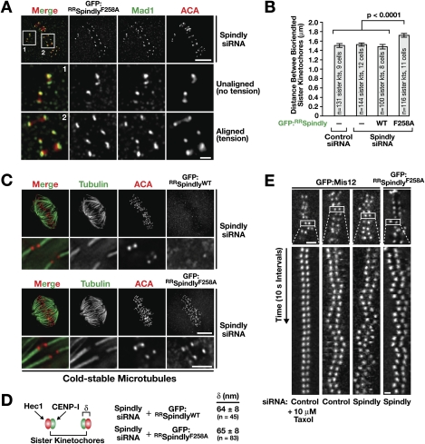
Figure 5.
Aligned kinetochores retaining Spindly motif mutants have achieved stable, bioriented microtubule attachments. (A) Projection of three optical sections from an immunofluorescence Z-stack of a cell depleted of endogenous Spindly expressing GFP:RRSpindlyF258A stained for GFP and Mad1. Cells were treated with siRNAs for 32 h, and expression of the Spindly transgenes was induced for 16 h before fixation. Bar, 5 μm; inset, 1 μm. (B) Distance between the ACA signal of sister kinetochores at metaphase in the indicated states. Error bars represent the SEM with a 95% confidence interval. (C) Cold-stable kinetochore fibers visualized by immunofluorescence in cells depleted of endogenous Spindly expressing GFP:RRSpindlyWT or GFP:RRSpindlyF258A. Blowups of individual kinetochore fibers represent projections of selected sections of the image Z-stack. Bar, 5 μm; blowups, 2 μm. (D) Distance (δ) between the inner kinetochore component CENP-I and the outer kinetochore component Ndc80/Hec1, measured at bioriented metaphase kinetochores in the indicated states. The value for unperturbed metaphase kinetochores in HeLa cells was previously determined to be 62 ± 9 nm (Wan et al. 2009). Values are given as the mean ± standard deviation. (E) Kymographs of aligned sister kinetochore pairs marked by GFP:Mis12 (Kline et al. 2006) or GFP:RRSpindlyF258A after the indicated treatments. Bar, 2 μm; kymograph panels, 1 μm.

