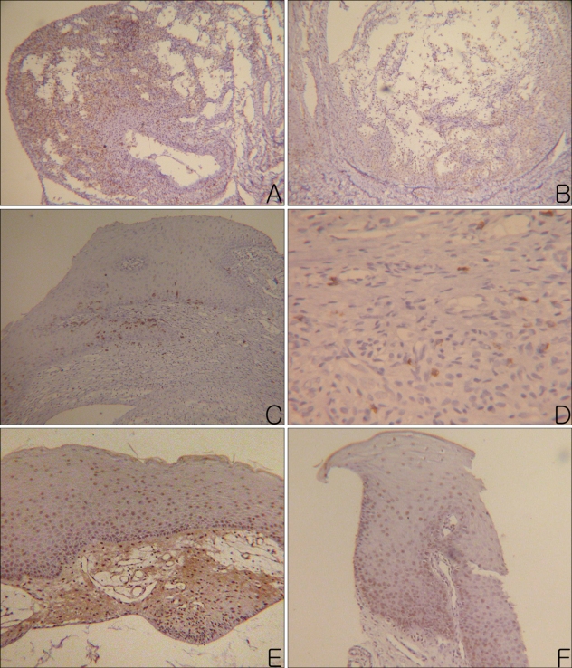
Fig. 3.
(A) Many CD4+ lymphocytes were observed in the granulation tissue of a mucocele. (B) Moderate number of CD4+ lymphocytes were observed in the wall and cavity of a mucocele. (C) Moderate numbers of CD8+ lymphocytes were observed in the mucosal basal layer and lamina propria. (D) A few CD8+ lymphocytes were observed in the granulation tissue, which formed the wall of a mucocele. (E) Apoptotic mucosal keratinocytes were observed evenly in the whole layer of the mucosa. (F) Apoptotic mucosal keratinocytes were observed especially on the basal layer of the mucosa (Left column photographs were taken from the PG and right column photographs were taken from NG. A, B: CD4, PAP, ×100, C: CD8, PAP, ×100, D: CD8, PAP, ×400, E, F: apoptosis, PAP, ×100).

