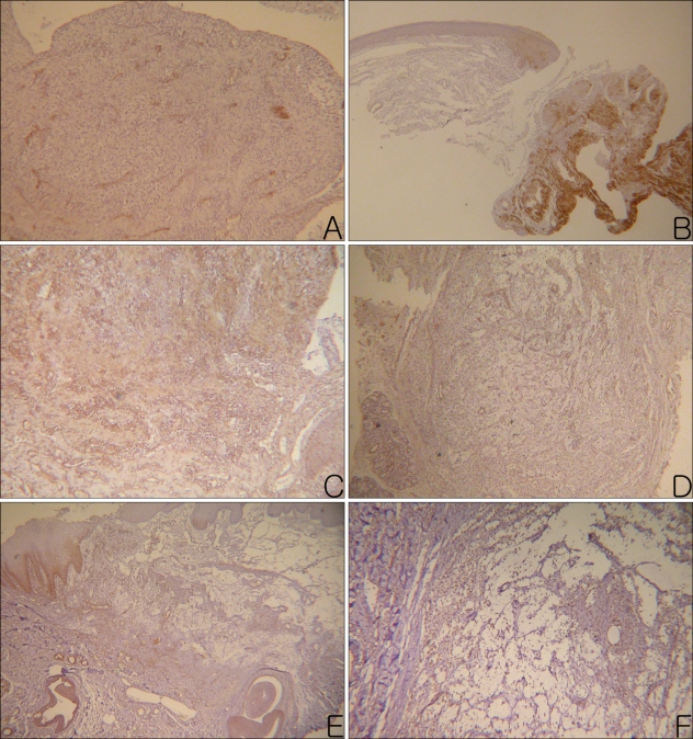
Fig. 4.
MMP-2+ materials (A, B), MMP-9+ materials (C, D), and TNF-α+ materials (E, F) were observed in the intercellular spaces of the granulation tissues and cavities of the mucoceles (Left column photographs were taken from PG and right column photographs were taken from the NG. A: MMP-2, PAP, ×100, B: MMP-2, PAP, ×40, C, D: MMP-9, PAP, ×100, E: TNF-α, PAP, ×40, F: TNF-α, PAP, ×200)

