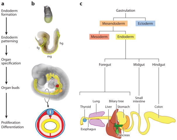
Figure 1.
Overview and timeline of endoderm organ formation. (a) The major events in endoderm organ formation are listed in chronological order and (b) illustrated with images of mouse embryos at e7.5 (top), e8.5, and e9.5 of development, with the endoderm shaded in (yellow). A schematic of a cross section through the e9.5 embryo illustrates the characteristic arrangement of the germ layers with the endoderm lining the gut tube (yellow), surrounded by mesoderm (red), and ectoderm (blue). (c) Endoderm cell lineages projected on to a schematic of the gastrointestinal tract. Fg; foregut, mg; midgut and hg; hindgut. The mouse embryo images are modified with permission from Zorn (2008).

