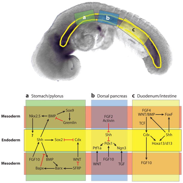
Figure 9.
Reciprocal epithelial-mesenchymal signaling in the gut tube during development of the stomach (a), pancreas (b), and duodenum/small intestine (c). The (upper panel) shows a 10-somite stage mouse embryo with the gut tube outlined in (yellow). The three boxes indicate the regions of the gut that give rise to the stomach (a), pancreas (b), and duodenum (c). The (lower panel) schematically shows these regions with the endoderm-epitheium (yellow) flanked by adjacent mesoderm-mesenchyme (above and below) (light red). The mesoderm was divided to allow for a summary of the multiple signaling cascades that have been identified in different species and at different stages of development. This schematic broadly summarizes our current understanding. In some cases the molecular relationships depicted have been directly demonstrated, whereas in other cases, we have inferred a connection from studies in other species.

