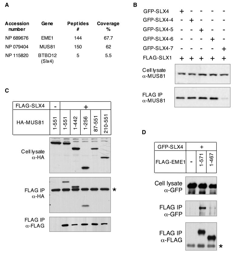
FIGURE 6. SLX4 associates with MUS81-EME1.
A- Table summarizing the number of peptides and the percentage of coverage obtained by Mass Spectrometric analysis of a FLAG-EME1 precipitate.
B- Western blot detection of endogenous MUS81 in total cell lysates of HEK cells co-producing FLAG-SLX1 and GFP-tagged full length or C-terminal SLX4 fragments and the corresponding FLAG-eluates. These are the same total cell lysates and FLAG-eluates than the ones described in Figure S10. The FLAG-eluates were also used in the nuclease assay shown in Figure 4E. The amount of total cell lysate loaded per lane is 10% of the input relative to the amount loaded of the corresponding FLAG-eluate.
C- and D- HEK cells were transfected with pcDNA3 constructs expressing the indicated deletion constructs of MUS81 or EME1 and full length SLX4. Cell lysate and α-FLAG immunoprecipitates (IP) were analyzed by western blot with the indicated antibodies.

