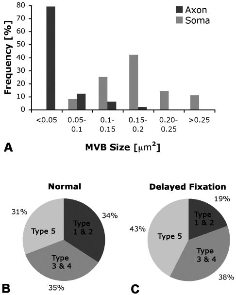
Fig. 3A-C. Quantification of MVB size and fractional area (FA) of five MVB types in hypoglossal neurons.
A. In axons, most MVBs are small, < 0.05 μm2, while most MVBs in the soma are 3-4 times larger, 0.1-0.2 μm2. B. In normal axons, small MVBs (types 1 and 2), classic (types 3 and 4), and late-endosomal type MVBs (type 5) make up a similar fraction (about one third) of the total fractional area (FA) of MVBs. C. With delayed fixation, the FA of small MVBs, types 1 and 2, decreases, while the FA of large MVBs, especially the late endosomal type 5, increases.

