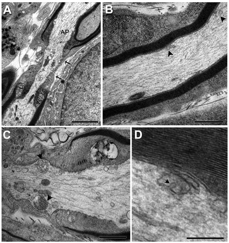
Fig. 4A-D. Morphological features of MVBs at the node of Ranvier and in the axon shaft of the hypoglossal nerve.
A. Representative section showing the axon at the node of Ranvier. The node is characterized by a lack of myelin and significantly restricted area of axoplasm (AP) in the “bottleneck” region (between arrows). B. High magnification of the axon shaft, characterized by dense myelin layers. Organelles (arrowheads) in the axon shaft are less numerous than in the paranode/node region. C. High magnification of the node of Ranvier. Note two MVBs (arrowheads), type 3 and type 4, in near symmetric positions, as well as numerous other organelles. D. High magnification of multivesicular-like organelle in an axon shaft apparently caught during membrane invagination (arrowhead) and formation of an internal vesicle. Scale bars: A = 2 μm, B and C =1 μm, D = 250 nm.

