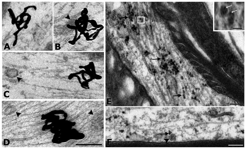
Fig. 7A-E. Retrogradely transported neurotrophic factors are not associated with MVBs in hypoglossal axons.
I125-GDNF and I125-BDNF were visualized by autoradiography of thin sections coated with a monolayer emulsion (A-D). A. Silver grain (SG) representing I125-GDNF, in axoplasm is not associated with any organelle. B. SG representing I125-GDNF in axoplasm next to an undefined structure (arrowhead). C. SG, representing I125-BDNF, is not associated with MVB type 1 (arrowhead). D. Dense SG in axoplasm is near, but not associated with MVB-like organelles (arrowheads). Quantum dot-conjugated BDNF (QD-BDNF) was evident by intense electron density of QDs in thin sections (E-F). E. Example of QD-BDNF in the node of a heavily-labeled axon. Many QDs appear to be associated with microtubules (arrows). At higher magnification (inset) it is apparent that the uniformly sized QD-BDNF is composed of multiple QDs (white arrows), possibly within a small endosomal organelle. Scale bar for inset = 50 nm. F. QD-conjugated BDNF (arrow) is located in close vicinity, but not within a “miniature” (type 1) MVB (arrowhead). Scale bars (same for panels A-D) = 200 nm.

