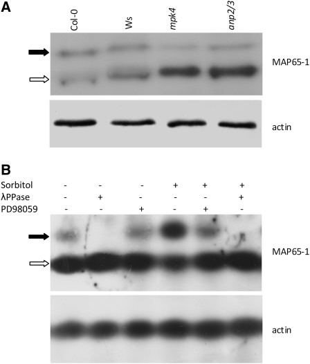
Figure 10.
Analysis of the Phosphorylation Status of MAP65-1 Using Phos-Tag.
(A) Phos-Tag SDS-PAGE coupled to immunoblot detection using a MAP65-1–specific antibody. The black arrow indicates the phosphorylated form (top bands retarded in mobility), while the white arrow indicates the nonphosphorylated form (bottom bands). In both the anp2 anp3 and the mpk4 mutants, the relative amount of the phospho-MAP65-1 form was dramatically reduced compared with the total amount of the protein. Anti-actin staining was used as a loading control.
(B) Probing the efficiency of Phos-Tag technology to discriminate phosphorylated from nonphosphorylated MAP65-1 in Col-0 extracts subjected or not to hyperosmotic stress (500 mM sorbitol, 30 min) in the absence or presence of the MAPKK inhibitor PD98059 (20 μM, 2 h pretreatment followed by 30 min with 500 mM sorbitol). The bottom band (white arrow) represents the nonphosphorylated form of MAP65-1. The top band (black arrow) represents the phosphorylated form of MAP65-1, which diminished during the PD98059 treatment and was completely eliminated following λ-phosphatase (λPPase) treatment of the extract prior to electrophoresis.

