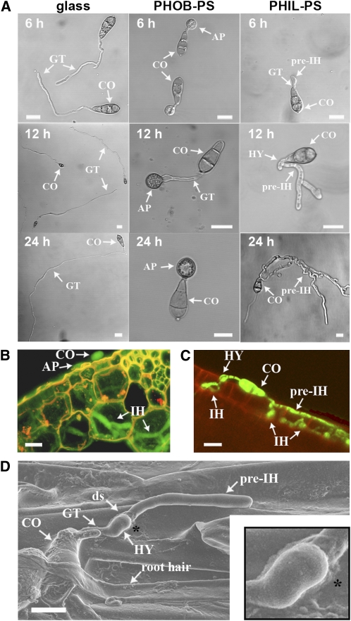
Figure 1.
Characterization of M. oryzae Development on PHIL-PS Surfaces and Roots.
(A) Time-course images of M. oryzae conidia (CO) germinated on different surfaces: glass, untreated polystyrene (PHOB-PS), or hydrophilic polystyrene (PHIL-PS). AP, appressoria; ds, developmental switch; GT, germ tubes; HY, hyphopodia.
(B) Cross section of a leaf sheath (plant cell wall autofluorescence in yellow) infected with a GFP-tagged M. oryzae strain (shown in green) in which IH can be seen.
(C) A GFP-tagged M. oryzae conidium (shown in green) on a root surface (plant cell wall autofluorescence in red); hyphopodia are observed on the surface and IH can be seen emerging from pre-IH within epidermal cells.
(D) Scanning electron micrograph of a germinating conidium growing on a root surface. A magnification of the developmental transition from germ tube to pre-IH (asterisk) is inset in the right-hand corner. ds, developmental switch.
Bars = 10 μm.

