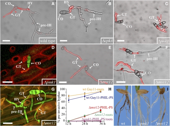
Figure 3.
Development of M. oryzae Mutants on Roots and Artificial Surfaces.
(A) to (B) M. oryzae wild-type strain Guy11 (A) and the Δcpka mutant (B) on hydrophilic polystyrene (PHIL-PS).
(C) and (D) Δpmk1 germinating conidia (CO) grown on either PHIL-PS (C) or on roots (D).
(E) to (G) Δmst12 mutant on either PHIL-PS ([E] and [F]), on which both types of growth (with and without pre-IH formation) are visualized, or on roots (G).
(H) Plot of time versus mean percentage of conidia of wild-type strain (diamonds), Δmst12 mutant (squares), and Δpmk1 mutant (triangles) developing pre-IH on PHIL-PS and roots (mean ± sd; n = 600 on PHIL-PS and 41 to 81 on roots; three experiments).
(I) Rice roots infected with wild-type strain or with Δpmk1 and Δmst12 mutants.
Fungal cell walls in (A) to (C), (E), and (F) show chitin content in red by TRITC-WGA fluorescence. CO, conidia; GT, germ tubes; HY, hyphopodia. Bars = 20 μm.

