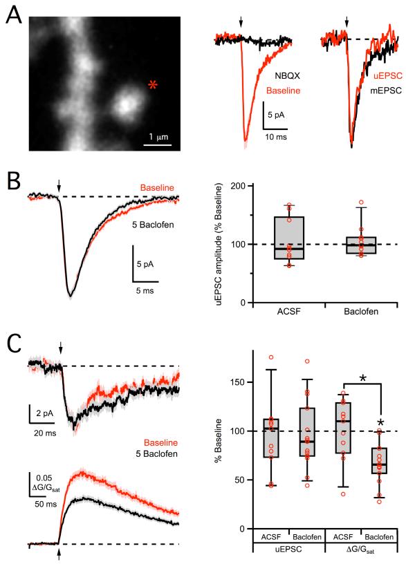
Figure 8. No modulation of AMPA-R or NMDA-R uEPSCs.
A, Left, 2PLSM image of dendrite and spine, showing the uncaging location (star). Middle, 2PLU (arrow) evokes an AMPA-R uEPSC at −70 mV (red) that is blocked by wash-in of NBQX (black). Right, Average AMPA-R uEPSC (red) is similar to average miniature EPSC recorded in the same cell (black). B, Left, Average AMPA-R uEPSCs in baseline conditions (red) and following wash-in of 5 μM baclofen (black). Right, Summary of changes in AMPA-R uEPSC amplitude following wash-in of baclofen or ACSF. C, Left, Average NMDA-R uEPSCs (top) and Ca signals (bottom), recorded at −70 mV in 0 mM extracellular Mg, in baseline conditions (red) and following wash-in of 5 μM baclofen (black). Right, Summary of changes in NMDA-R uEPSC and Ca signal amplitudes following wash-in of baclofen or ACSF. Asterisks indicate significant (P < 0.05) difference from 100% or between different conditions. See also Figure S3.

