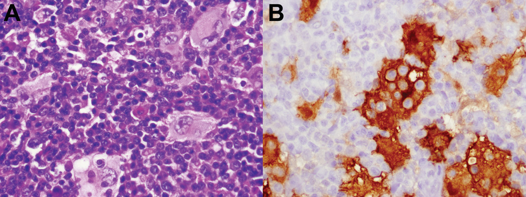
Figure 3.
Rosai-Dorfman disease, axillary node biopsy, age 9. A. Characteristic histiocytes with abundant pale cytoplasm show emperipolesis. B. Histiocytes are strongly S100-positive, highlighting engulfed lymphoid cells. (S100 immunoperoxidase). They were negative for CD1a, CD21 and langerin. (not shown)

