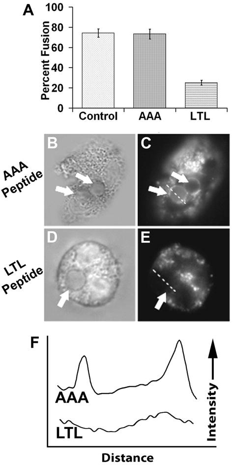
Fig. 6.

Trojan-LTL peptide treatment blocks phagosome-lysosome fusion. Adherent neutrophils were treated with the Trojan-AAA or Trojan-LTL peptides then allowed to internalize EA. After a two hour incubation, cells were labeled with LysoTracker and imaged as described in the Materials and Methods. Phagosomes were scored for phagosome-lysosome fusion. The percentage of labeled phagosomes (phagolysosomes) was tallied for the various cell treatments (A). Untreated cells (control) and cells treated with the Trojan-AAA peptide show indistinguishable fusion percentages, while LTL treated cells show a much lower percentage of phagolysosome formation (p<0.0001). Panels B–E show examples of lysosome labeling during Trojan-AAA (B and C) and Trojan-LTL (D and E) treatment. Phagosomes are denoted with arrows. Lysosome-fused phagosmes in C have a distinctive bright ring around the edge, whereas the unfused phagosome in E has no apparent labeling. Dashed lines through phagosomes in C and E show the line used for line profile analyses (F, traces AAA and LTL, respectively). The line profiles show plots of LysoTracker intensity across the distance of the line shown in the corresponding micrograph. (n=4) (A, error bars represent standard deviation; B–E, x1400)
