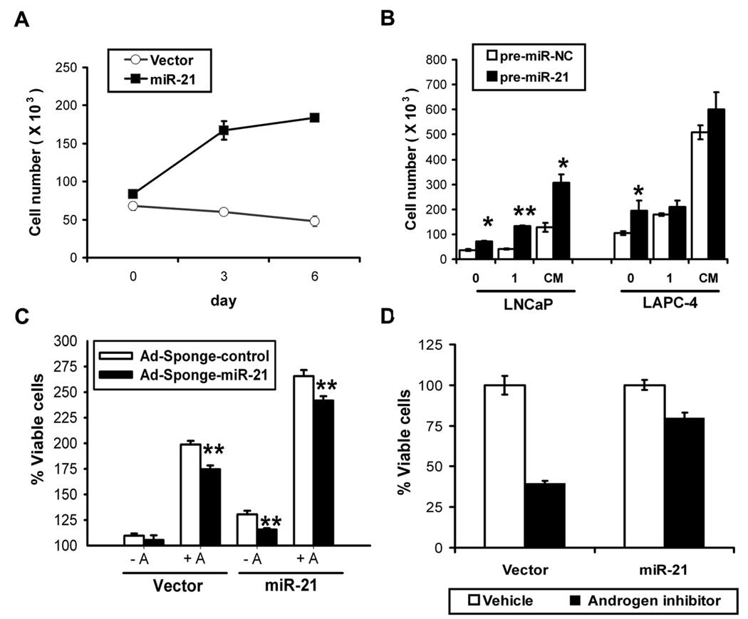
Fig. 2. miR-21-induced proliferation.
A, miR-21 mediated androgen-independent growth. LNCaP retrovirally transduced to stably express miR-21 (black square) or empty vector (white circle) grown in androgen-depleted media. Mean ± S.E. B, Effects of synthetic miR-21 on growth. Transient transfectantions with synthetic pre-miR-21 (black) or pre-miR-Negative Control (white) after 6 days in androgen-depleted (0), 1 nM R1881 (1) or complete media (CM). Viable cells quantified by trypan blue (Mean ± S.E. from 3 different wells), *P<0.05, **P<0.01 (t-student test). C, Effects of miR-21 inhibition. LNCaP miR-21 or vector control cells infected with 10 MOI of Ad-Sponge-control (white) or anti-miR-21 Ad-Sponge-miR-21 (black) after 6 days in charcoal-stripped (−A) or R1881 supplemented (+A) media. Percent growth calculated by MTS. Mean ± S.E of 12 independent measurements, **P<0.01 (t-student test). D, Elevated miR-21 partially overcomes AR blockade. LNCaP miR-21 or control vector in complete media treated with vehicle (white) or 10 µM Casodex (black). Cell proliferation calculated by MTS (relative to vehicle, 6 days). Mean ± S.E of 6 independent measurements.

