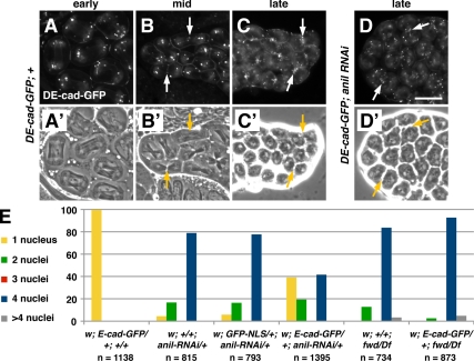
Figure 6.
DE-cad-GFP suppresses cytokinesis defects caused by anillin depletion. (A–D′) Fluorescence (A–D) and corresponding phase-contrast (A′–D′) micrographs of dividing spermatoctyes expressing DE-cad-GFP. (A and A′) In wild-type cells DE-cad-GFP localizes to the cortex and in puncta at the poles during anaphase. During telophase, DE-cad-GFP begins to accumulate at the equator (arrows; B and B′). By late telophase, DE-cad-GFP becomes highly concentrated in the furrow in wild-type (C and C′) and dsRNA-expressing cells (D and D′). (E) Expression of DE-cad-GFP greatly reduces the percentage of multinucleate spermatids in flies expressing dsRNA directed against anillin but not in flies mutant for fwd. The number of spermatids counted for each genotype is indicated (n). Note that although these results were obtained with the UAS::anillin-RNAi line, similar results were obtained for β2t::anillin-RNAi (not shown; see Materials and Methods). Bar, 20 μm.

