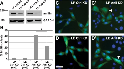
Figure 9.
Expression of E-cadherin suppresses cytokinesis defects caused by depletion of anillin in mouse L-cells. (A) Immunoblots showing reduced levels of anillin protein (∼190 kDa) in anillin shRNA-expressing mouse LP (empty vector) or LE (E-cadherin expressing) cells (Anil KD) compared with scrambled shRNA-expressing cells (Ctrl KD). GAPDH (∼40 kDa) is the loading control. (B) Quantitation of multinucleate cells confirms suppression of anillin loss by expression of E-cadherin. *p = 0.00076. n is the number of experiments, and 100 cells were counted in triplicate for each experiment. (C and D) Micrographs of LP and LE cells expressing scrambled shRNA (C and D) or anillin shRNA (C′ and D′) marked by GFP (green). DNA is stained with Hoechst (blue). Representative multinucleate cells are marked (white arrowheads). Bar, 100 μm.

