Figure 4.
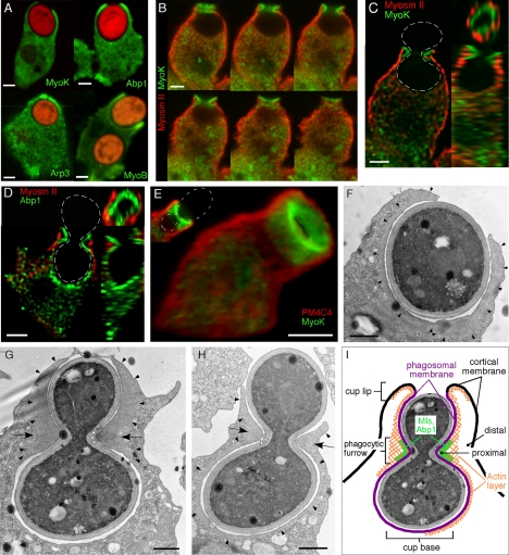
Enrichment of cytoskeletal proteins in the phagocytic cup and phagocytic furrow. (A) MyoK, Abp1, Arp3, and MyoB are enriched at the lip of the phagocytic cup. Cells were fed with TRITC-labeled fluorescent yeasts for 5 min. MyoK, Abp1, and Arp3 were localized by specific antibody detection in MyoK-overexpressing cells (MyoK OE) or wild-type cells, respectively. The GFP-MyoB signal was enhanced by anti-GFP antibodies. (B–E) Enrichment of MyoK and Abp1 around the bud neck of engulfed yeast defines the phagocytic furrow. Dotted white lines represent the contour of budded yeasts. (B) Serial averaged projections of three confocal sections of cells immunostained for MyoK and myosin II. (C) xy, yz, and xz plane sections of the series shown in B. (D) xy, yz, and xz plane sections of a cell immunostained for Abp1 and myosin II. (E) 3D reconstruction of a phagocytic furrow stained for MyoK and PM4C4. A single section of the same image is presented (top left corner). (F–H) Electron micrographs of phagocytosed yeasts in wild-type cells. (F) Phagocytic cup enclosing single yeast displayed actin enrichment not only around the phagosomal membrane (closed arrowheads) but also lining the cortical plasma membrane near the lip (open arrowheads). (G and H) Phagocytic cups enclosing budded yeasts at two stages of closure. Arrows point at the actin-rich zone of the phagocytic furrow. A denser dark area in the actin-rich zone of the furrow (open arrowheads) is particularly visible in G. The actin layer surrounding the cup is continuous with the actin-rich zone in the furrow (closed arrowheads). This actin layer is visible at the lip in G and H, whereas it is absent from the cup base in G. (I) Schematic representation of the phagocytic cup with a furrow and definition of the terms used in this study. Bars, 2 μm (A–E) or 1 μm (F–H).

