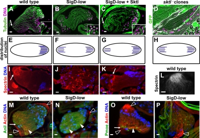
Figure 3.
Normal levels of PIP2 are required for spermatid cyst polarity and polarized distribution of F-actin and actin-associated proteins. Fluorescence micrographs of elongating spermatid cysts. Arrows, growing ends. (A–C) Confocal micrographs of elongated Drosophila cysts stained for DNA (magenta) and tubulin (green). (A) Wild-type cyst with nuclei at one end. (B) SigD-low bipolar cyst, with nuclei at both ends. (C) Coexpression of Sktl with SigD-low partially rescues cyst polarity. (Insets, A–C) Single confocal sections showing perinuclear microtubule arrays, which are disorganized in SigD-low. Tubulin was stained with antibodies directed against acetylated α-tubulin (A and C) or GFP (to visualize β-tubulin-GFP (B). (D) Epifluorescence micrograph of a live squashed preparation of male germ cells stained for DNA (magenta), showing a bipolar sktl2.3 mutant clone (see Materials and Methods). Cysts mutant for sktl2.3 are marked by the absence of GFP (green). (E–H) Diagrams illustrating the polarity of wild-type (E), SigD-low (F), partially rescued (SigD-low + Sktl; G) and sktl− (H) cysts. (I–K) Epifluorescence micrographs showing elongated spermatid cysts stained for α-spectrin (red) and DNA (blue). In wild type (I), spectrin localizes to the growing end of the elongating cyst. In SigD-low (J), spectrin localizes to the middle of the cyst. (K) Localization of spectrin is partially rescued by coexpression of Sktl with SigD-low. (L) Confocal image showing localization of α-spectrin in the honeycomb at the growing end of a wild-type cyst. (M–P) Epifluorescence micrographs of elongating cysts showing F-actin (red), DNA (blue), and anillin (Anil, green), and M and P-moesin (Pmoe, green; O and P). Colocalization (yellow). In wild type, anillin (M) and P-moesin (O) localize to ring canals near where F-actin is concentrated at the growing end (arrowheads, arrows). In SigD-low, anillin (N) and P-moesin (P) remain associated with ring canals, which are scattered (N) or which localize to the middle of the cyst (P). Actin filaments are distributed along the cyst (arrowheads, N). White open arrowheads, cyst cells. Scale bars (A–C and L–P), 10 μm; (D and I–K) 20 μm.

