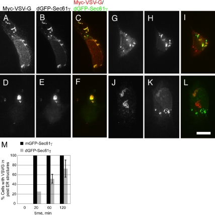
Figure 9.
ER membrane stacking is sufficient to slow ER export. Cells cotransfected with Myc-ts045 VSV-G and either a control mGFP-Sec61γ construct (not shown but quantified in M) or the ER stack-inducing dGFP-Sec61γ construct were shifted to 40°C to accumulate VSV-G in the ER. Thereafter, cells were shifted to 32°C for 0 (A–C), 20 (D–F), 60 (G–I), or 120 (J–L) min. At the indicated times, cells were fixed and stained with Myc epitope antibodies. The corresponding VSV-G (A, D, G, and J) and GFP-Sec61γ (B, E, H, and K) as well as merged images (C, F, I, and L) are shown. Bar, 10 μm. (M) Quantitation of the kinetics of VSV-G export under each condition, the average of two independent experiments, ±SD is shown.

