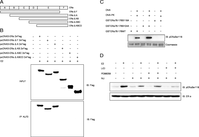
Figure 2.
Ku70 interacts with the B domain of ERα. (A) ERα deletion mutants generated by PCR. (B) COS-7 cells were cotransfected with Flag-tagged wild-type ERα or ERα deletion mutants (ΔF, ΔA, ΔAB, ΔABC, and ΔABCD). Forty-eight hours after transfection, cells were lysed, and Ku70 was immunoprecipitated with anti-Ku70. The lysates (top) and immunoprecipitates (IP; bottom) were immunoblotted (IB) with anti-Flag M2 antibody. (C) In vitro kinase assay using wild-type and mutant GST-ERα-(76-176) fusion proteins (1 μg) as substrates for DNA-PK (top, pERαSer118; bottom, Coomassie staining). (D) MELN cells were incubated for 10 min with 10 mM LiCl, 5 μM NU7026, or 10 μM PD98059 and thereafter with 100 nM E2 for 20 min. ERα phosphorylation was detected by immunoblotting. Detection of β-actin was used as a loading control.

