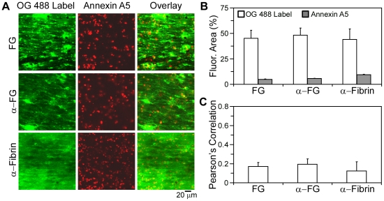
Figure 4. Spatial localization of fibrin(ogen) and procoagulant platelets during thrombus formation.
Thrombi were formed by a 15 minute co-perfusion of whole blood and CaCl2/TF at 200 s−1 over fibrinogen. Dual-labeling was with AF647-annexin A5 (14 nmol/L, red) and the indicated fibrin(ogen) labels (green). A, Confocal fluorescence images in green are shown for OG488-fibrinogen (FG, 0.3 µmol/L), anti-fibrinogen antibody (α-FG, 20 µg/mL), or anti-fibrin antibody (α-Fibrin, 20 µg/mL). Images in red indicate staining of PS-exposing platelets. B, Fluorescence surface area coverage for each OG488 label and annexin A5. C, Pearson's correlation coefficient (Rr), between annexin A5 and each green label. Mean ± SEM (n = 3–5 experiments).

