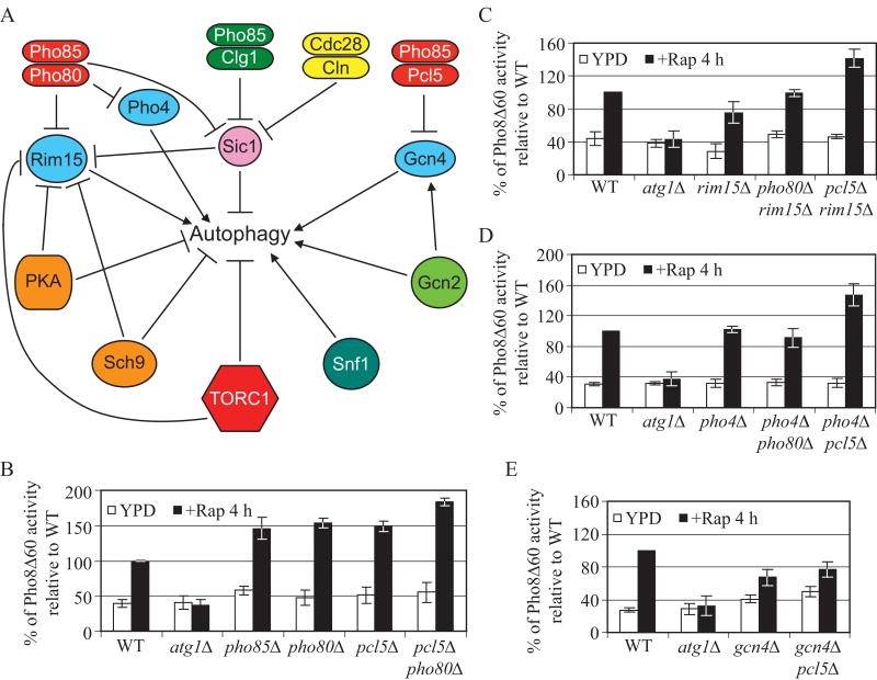
Figure 1.
Pho80 and Pcl5 are the cyclins of Pho85 that participate in the negative regulation of autophagy.
(A) Schematic overview of the key components in autophagy regulation. Arrows represent positive regulation; bars represent negative regulation. Both Clg1 and Pcl1 (not shown) are cyclins that form positive regulatory complexes with Pho85 through the inhibition of Sic1.
(B), (C), (D) and (E) Cells expressing Pho8Δ60 were grown in YPD to midlog phase and then treated with rapamycin for 4 h. The Pho8Δ60 activity was measured as described in Supplemental Experimental Procedures, and was normalized to the activity of wild-type cells with rapamycin treatment, which was set to 100%. Error bars indicate the standard deviation (SD) of three independent experiments. Strains used were wild type (TN124), and atg1Δ (HAY572), and in (B) pho85Δ (ZFY089), pho80Δ (ZFY105), pcl5Δ (ZFY099) and pcl5Δ pho80Δ (ZFY128); in (C) rim15Δ (ZFY100), pho80Δ rim15Δ (ZFY102) and pcl5Δ rim15Δ (ZFY103); in (D) pho4Δ (ZFY135), pho4Δ pho80Δ (ZFY137) and pcl5Δ pho4Δ (ZFY143); in (E) gcn4Δ (ZFY111), gcn4Δ pcl5Δ (ZFY112). See also Figure S1.

