
Figure 2.
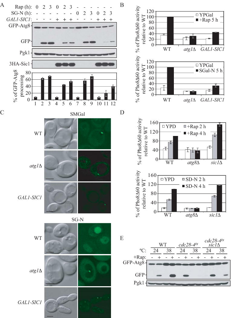
Sic1 functions as a negative regulator of autophagy.
(A) and (B) Overexpression of Sic1 inhibits rapamycin- and nitrogen starvation-induced autophagy. (A) Wild-type (W303-1B) cells expressing GFP-Atg8 (pCU-GFP-AUT7(414)) and expressing either 3HA-Sic1 (pZY011), or an empty vector (pTY006), were grown in SMD and shifted to SMGal for 12 h, and then subjected to either rapamycin treatment or starvation treatment (SG-N). At the indicated times, proteins were TCA-precipitated and subjected to immunoblotting with anti-YFP, anti-HA and anti-Pgk1 (loading control) antisera. Percentage of GFP-Atg8 processing was calculated as described in Supplemental Experimental Procedures. Error bars indicate the SD of three independent experiments.
(B) Wild-type (ZFY202), atg1Δ (TYY181) and GAL1-SIC1 (ZFY203) cells expressing Pho8Δ60 were grown in YPD and shifted to YPGal for 12 h, and were then treated with rapamycin or shifted to nitrogen starvation (SG-N) medium for 5 h. The Pho8Δ60 activity was normalized to the activity of wild-type cells treated with rapamycin or nitrogen starved, which was set to 100%. Error bars indicate the SD of three independent experiments.
(C) Overexpression of Sic1 inhibits delivery of GFP-Atg8 to the vacuole. Wild type (W303-1B), atg1Δ (TYY164), and GAL1-SIC1 (ZFY184) cells expressing GFP-Atg8 (pCU-GFP-AUT7(416)), were analyzed by fluorescence microscopy as described in Supplemental Experimental Procedures. Bar, 5 μm.
(D) Nonspecific autophagy is elevated upon deletion of SIC1. Wild-type (TN124), atg8Δ (YZX200) and sic1Δ (ZFY098) cells expressing Pho8Δ60 were grown to midlog phase and treated with rapamycin, or shifted to nitrogen starvation conditions (SD-N). At the indicated times, the Pho8Δ60 activity was measured, and it was normalized to the activity of wild-type cells with nitrogen starvation or rapamycin treatment for 4 h, which was set to 100%. Error bars indicate the SD of three independent experiments.
(E) The autophagic defect in the cdc28-4ts mutant is partially suppressed by deletion of SIC1. Wild-type cells (BY4742) and temperature-sensitive mutants cdc28-4ts (D4), and cdc28-4ts sic1Δ (ZFY258) expressing GFP-Atg8 (pCU-GFP-AUT7(416)), were grown at 24°C or 38°C for 3 h, and treated with rapamycin for 2 h. TCA-precipitated proteins were subjected to immunoblotting with anti-YFP and anti-Pgk1 (loading control) antisera. See also Figure S2.