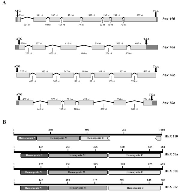
Figure 1.
Hexamerin genes and deduced protein subunits. (A) Schematic diagram of hexamerin genes structure. Exons and introns are represented by boxes and lines, respectively. Arrows at the right and left of each gene indicate initiation and termination codons, respectively. Sequenced untranslated regions (UTR) are marked in dark gray. Number of nucleotides (nt) are indicated for exons and introns (exons and the primers used for hexamerin sequencing are marked in the nucleotide sequences shown in Additional files 1, 2, 3 and 4). (B) Diagrams of the deduced hexamerin proteins showing the N, M and C hemocyanin domains. Note that the C domain is interrupted in the HEX 110 sequence. Signal peptides and conserved motifs are marked in hexamerin sequences shown in Additional files 5, 6, 7 and 8.

