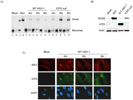
Figure 1. ICP0 inhibits the sustained activation of IRF3 during the later stages of an HSV-1 infection.
(A) HEL cells were mock treated, infected with WT HSV-1 (strain F) or a corresponding ICP0 null virus (R7910). Cytoplasmic and nuclear protein extracts were resolved using native western blotting to assess IRF3 dimerization. (B) Western blot analysis of whole cell protein lysates collected from HEL cells after 8 hours of infection as indicated. (C) Immunofluorescence microscopy examining ICP0 and IRF3 subcellular localization following a time course infection of HEL cells with WT HSV-1. Cell nuclei were hoechst stained (DAPI). A representative field of view is shown for mock treated cells. In all cases, SeV served as a positive control for activation of the IRF3 pathway.

