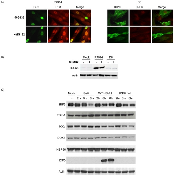
Figure 6. Proteasome activity facilitates appropriate subcellular localization of ICP0 and not degradation of IRF3 constituents.
(A) Immunofluorescence microscopy was used to examine the localization of ICP0 and IRF3 in HEL cells following an 8 hour infection with R7914 or D8 in the absence or presence of MG132. (B) HEL cells were treated as described in part A and ISG56 expression was assessed by western blot analysis. (C) The expression of IRF3 pathway constituents was examined by western blot analysis following a time course infection of HEL cells with WT HSV-1 (strain F) or an ICP0 null virus (R7910). SeV was employed as a positive control for the activation of IRF3.

