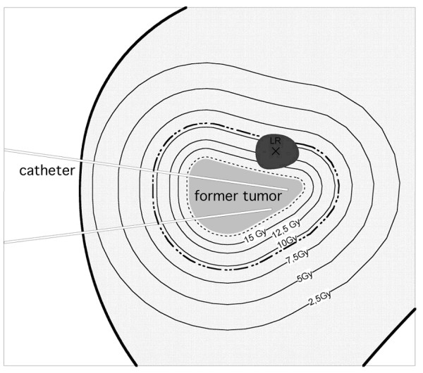
Figure 1.

Scheme of a follow-up MRI merged with the initial dosimetry displaying a tumor recurrence of a micrometastasis (LR). The black cross in LR marks the 3D isocenter. The dashed line describes the CTV around the colorectal liver metastasis which had been treated initially. The bold dashed line outlines 23.5 mm distance from the initial tumor margin.
