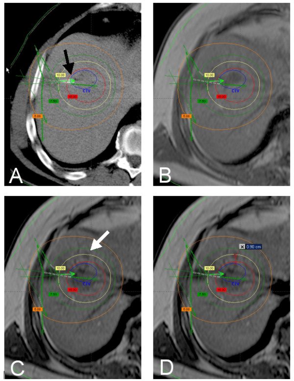
Figure 2.

A: planning CT with overlayed dosimetry (BrachyVision®) showing a colorectal liver metastasis in segment 8. One catheter tip is displayed directly (black arrow), more catheters in other levels of the liver are indicated by green arrows. Verification of correct definition of the CTV was performed by image fusion of the planning CT with a MR scan (T1 GRE without contrast media) obtained 3 days prior to treatment (B). Local recurrence (white arrow) 6 months after treatment (MR, T1 GRE without contrast media, C). The distance of the 3D isocenter of the local recurrence from the initial tumor margin is 9 mm. Thus, the local tumor recurrence meets the criteria for micrometastasis growth (D). The dose initially applied in the center of the micrometastasis was 10.9 Gy.
