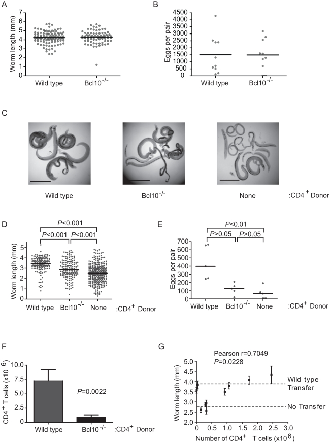
Figure 1. TCR signaling in CD4+ T cells is dispensable for S. mansoni development.
(A) Wild type and Bcl10-/- mice were infected with S. mansoni, parasites were recovered from the portal tract at 6 weeks post infection and male worms were measured from digital micrographs. Mean values are represented by horizontal bars. (B) Egg production by schistosome pairs in wild type and Bcl10-/- mice at 6 weeks post infection. Mean values are represented by horizontal bars. (C) RAG-1-/- mice reconstituted with wild type or Bcl10-/- CD4+ T cells mice were infected with S. mansoni and parasites were recovered from the portal tract at 6 weeks post infection. Representative micrographs of parasites recovered from non-reconstituted RAG-1-/- mice and RAG-1-/- mice reconstituted with wild type or Bcl10-/- CD4+ T cells are shown. Bar = 1 mm. (D) Lengths of male worms recovered from reconstituted RAG-1-/- mice described in (C) were measured from digital micrographs. Mean values are represented by horizontal bars. (E) Egg production by schistosome pairs in reconstituted RAG-1-/- mice described in (C) at 6 weeks post infection. Mean values are represented by horizontal bars. (F) At necropsy, the numbers of CD4+ T cells in the spleens of RAG-1-/- recipients were determined by flow cytometry. Data are represented as mean +/− SEM. (G) Correlation of average male worm length and CD4+ T cell numbers for each RAG-1-/- recipient of Bcl10-/- CD4+ T cells. “WT Transfer” and “No Transfer” dashed lines represent the average length of parasites recovered from control mice that received wild type CD4+ T cells or PBS alone, respectively. Worm length data for each mouse are represented as mean +/− SEM. Groups of 4–5 mice were used for each experimental condition. Data are representative of three independent experiments.

