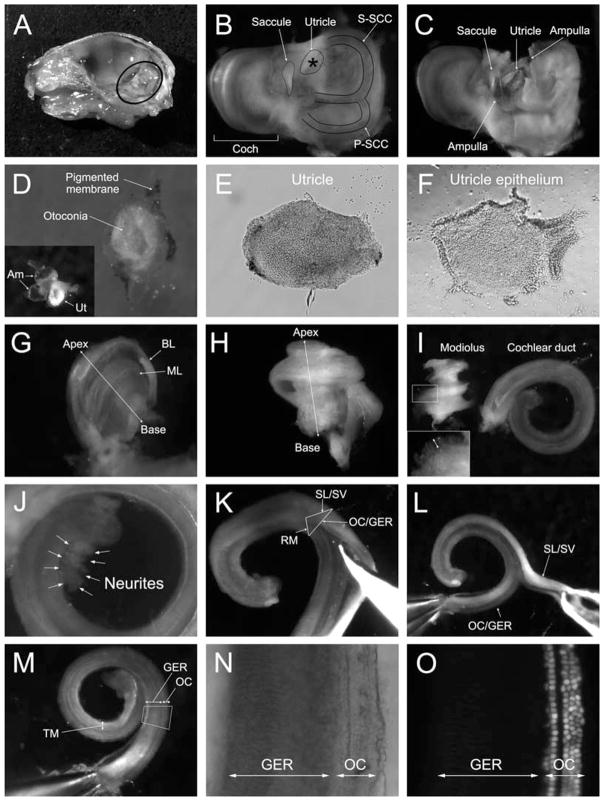
Fig. 9.1.
Dissection of utricle, cochlear duct, and modiolus. (A) Right side of a bisected skull of a 1-day-old (P1) mouse after removal of the brain. Circled area shows the petrous (temporal) bone containing the inner ear organs to be dissected. (B) The inner ear after removal of the bulla and surrounding tissue. The asterisk (*) indicates the cartilage overlying the utricle. Coch = cochlear part, S-SCC = superior semicircular canal, and P-SCC = posterior semicircular canal. (C) The utricle is exposed by fenestration of the overlying cartilaginous plate. (D) Dissected utricle. The pigmented membrane is pulled aside and mostly removed to expose the epithelium covered with white otoconia. The inset picture shows the utricle (Ut) with two ampullae (Am), which belong to the superior SCC and horizontal SCC. (E) The utricle after complete removal of the pigmented membrane and otoconia. (F) The utricle epithelium obtained from the same utricle as in E after thermolysin treatment. (G) The bony labyrinth (BL) of the cochlea is partially removed to expose the membranous labyrinth (ML) of the cochlea. (H) Side view of the whole membranous labyrinth of the cochlea. (I) The cochlear duct is peeled off from the modiolus. The tiny protrusions (arrow in the inset) around the modiolus are the neuronal fibers that innervated the hair cells. Note that there should be no neuronal tissue (neurites or cellular material) visibly attached to the duct. (J) Example of an unacceptable cochlear duct preparation. The arrows labeled “Neurites” indicate contaminating neuronal tissue attached to the duct. (K) Reissner’s membrane is cut with forceps from the most basal to the apical part to open the cochlear duct. The triangle illustrates the cross-section of the cochlear duct. RM = Reissner’s membrane, SL/SV = spiral ligament with stria vascularis, and OC/GER = the organ of Corti (OC) with the greater epithelial ridge (GER). (L) The spiral ligament and stria vascularis (SL/SV) are removed from the OC by carefully tearing the tissues apart. (M) View of the OC with the GER and tectorial membrane (TM). (N) A higher magnified image of the squared area in (M). OC indicates the organ of Corti and GER labels the greater epithelial ridge. (O) An epifluorescent image of (N) in which the nuclei of cochlear hair cells can be identified by their bright nuclear fluorescence. This nuclear green fluorescence of hair cells is a distinct feature of the Math1-nGFP transgenic mouse (16) used in the dissection shown.

