Fig. 9.3.
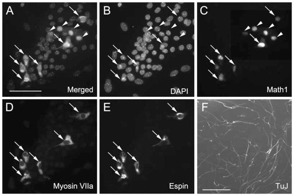
Differentiation of spheres derived from the utricle and spiral ganglion. (A–E) After a 2-week differentiation period, hair cell–like cells appear in some of the cells derived from utricle-derived spheres. (A) Merged image of (B–E). (B) All cell nuclei are visualized with DAPI. (C) Cells expressing nuclear green fluorescence as seen by bright spots that overlap with the nuclear staining shown in (B) are indicative of Atoh1 promoter activity in the Math1/nGFP mouse used for sphere preparation (16) (Math1). (D) Myosin VIIA immunoreactivity, and (E) immunostaining for espin expression. Arrows in (A–E) point to triple hair cell marker–positive cells expressing nGFP (Math-1), myosin VIIA, and espin. Arrowheads point to cells that are only positive for nGFP (Math-1) and negative for the other two markers, suggesting that these cells have not yet upregulated myosin VIIA and espin. (F) Cells that differentiated from spiral ganglion-derived spheres. After the 2-week differentiation period, (TuJ)-positive neuronal marker is detectable in cells with distinct neural morphology. Scale bar = 50 μm in (A–E) and 500 μm in (F).

