
Fig. 2.
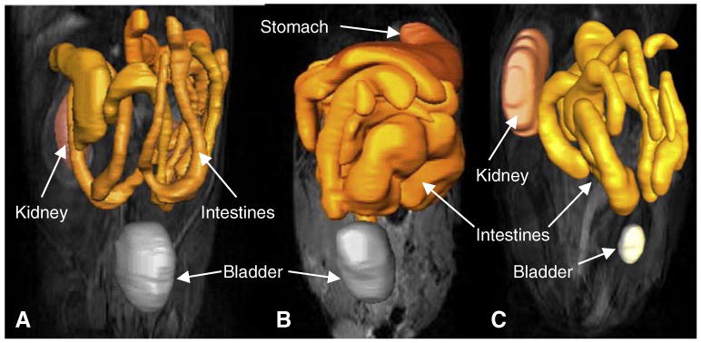
Representative MRI of uninfected (a), unsupplemented infected (b), and Se-supplemented infected (c) mice. Images were acquired of the entire mouse GI tract. 3D reconstruction of the GI tract was created and is overlayed on one of the MRI images. Enlargement of the intestines is observed in the infected mouse (b)