Fig. 6.
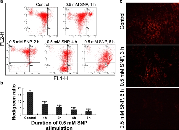
Sodium nitroprusside (SNP) treatment leads to a decrease in mitochondrial membrane potential. a Typical pictures output by a flow cytometer. Green FL1-H, red FL2-H. b Comparison of mitochondrial membrane potential (red/green ratio) between groups. As compared with the control, ***P < 0.001. c In situ JC-1 staining (original magnification, ×320). Decreased red fluorescence intensity indicating decreased amount of JC-1 polymer caused by decreased mitochondrial membrane potential

